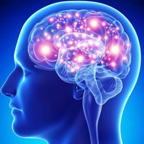
How can we start diagnostics of neurological diseases when still in an ambulance car, where there is no bulky medical equipment? And how can we determine if a patient has started to recover after a serious disease or brain injury?
Our scientists are developing a diagnostic system which in the future will allow conducting express screening of brain condition and transmit information to a medical centre.
This hardware and software mini complex excited big interest at Cognitive Neuroscience - 2018 International Forum on Cognitive Neurosciences which took place in Yekaterinburg. What can it offer for study of the brain and early diagnosis of its “concealed defects”? This became the main topic of discussion with one of the authors of the medical technology being developed, Anton Pashkov, postgraduate student of the SUSU Department of Clinical Psychology.
Express-diagnostician
— What inspired you to create the computer mini diagnostician?
— In order to recognize a stroke or other, frequently occult, latent brain disorders in a clinic, we have to use bulk stationary units, for example, computer or magnetic resonance imaging scanners. But they can’t be brought when visiting a patient at home. So our portable device on the basis of electroencephalography (EEG), which register electrical activity of the brain, will allow conducting this extremely difficult diagnostics even before patient’s arrival to a hospital. To do this, one should simply put on a “magic hat” with sensors.
— What are the advantages of your method?
— This is a completely new approach being implemented within development of telemedicine. One extra advantage of our method, compared to the MRI, is a higher time resolution. Thus, we simultaneously have several information sources which reflect various aspects of cerebral operation.
It should be noted that such large-scale projects require interdisciplinary participation. For example, software for the hardware and software complex is being developed in cooperation with the SUSU School of Electrical Engineering and Computer Science.
— So you are saying that it will be possible to recognize complex brain diseases right in the ambulance?
— Partly it is so. Our invention can become especially useful at initial stages of a patient’s transportation to the nearest medical institution and his or her subsequent hospitalization. This mobile complex is compact; it can fit into a portable satchel and it can be used in field conditions.
Moreover, within the Telemedicine project, we are developing a new system for EEG signal transmission via secured communication channel. Information will be streaming online to the nearest neurological centre. There it will be analysed by specialists, and by the time that a patient arrives to the hospital, specialists will already have the initial data which will help in further diagnostics and in choosing therapy or rehabilitation programme. We are getting ready for testing of the innovative system.
Without incision
— How can your technology help after treatment?
— It can be used to monitor condition of a patient during rehabilitation with therapeutic purposes. Modern innovative non-invasive (without incision) methods for assessment of efficiency of neurorehabilitation programmes allow constantly monitoring condition of a patient and adjust the chosen strategy of treatment based on these data.
— Will this technology get further development?
— We are planning to go further and integrate widely used methods of EEG data analysis with medical potential of one of the neuroscience’s fields which is trending nowadays, the so-called connectomics. This science studies totality of connections between neural regions of the brain.
For instance, we are planning to research topological properties of neural networks, for example, of the “small world”, which are an indicator of some balance between segregation (splitting) and integration of information provided by different regions of brain. In other words, it splits information at first in order to integrate it in one piece later. This provides new possibilities for disease diagnostics based on a search for biomarkers – physiologically and numerically measurable indicators of operation of certain organism systems, including the brain.
.jpg)
Biomarker of stress
— But this is only one of the biomarkers…
— Sure it is, but there are the others. Let us consider, for example, the network of passive operation mode of the brain. It characterizes the state when a person is calm and relaxed. This network was discovered in 2001 by professor Marcus Raichle from Washington University at the U.S. Any negative events – stresses, symptoms of depression, anxiety – get reflected in measurements of this neural network.
— So what does it mean?
— Aside from the fact that such biomarker can be very useful for diagnostics in a clinic, this allows us expanding theoretical knowledge about operation of human brain. Moreover, it is known nowadays that aside from the network of passive operation mode of the brain, there are more than 10 of such networks (they can be found using imaging), so we can track their interactions in both healthy people under test and in patients. We get a chance to use dynamic nature of such interactions for assessment of efficiency of neurorehabilitation programmes in a neurology clinic, as well as efficiency of teraphy with the use of antidepressants, neuroleptics and tranquilizers…
— Is it true that sleeping is healing?
— Being an inextricable part of our life, healthy sleep is one of the formulas for mental and somatic health. The study of physiological fundamentals of sleep is one of the key directions of both fundamental and applied research. As of today, we know that this is a universal phenomenon for many living organisms. Sleep, or more precisely, sleeplike conditions are observed among nematodes, reptiles, birds, and, of course, mammals. Nowadays, understanding the mechanisms of brain operation while sleeping is of a great interest for science. And our mini scanner of the brain provides new opportunities for such researchers.
— Is it true that brain cells can’t regenerate? Is there hope for their regeneration?
— This is a myth. Nerve cells can regenerate. Besides, human brain has two unique areas where neurons can originate throughout a person’s life: dentate gyrus and the subventicular zone. Under insignificant damage, neurons “repair” themselves due to their ability to reparation.
Virtual model
— Will the artificial intelligence be able to perform self-learning in the course of research?
— This is exactly what’s happening! Speaking the language of programmers, this is machine learning. Within Project 5-100, we start using artificial neural networks in order to perform analysis of EEG signals. We are “feeding” them with data about state of the brain of healthy and ill patients, and while comparing them, the machine can independently divide them into groups based on health condition.
These intelligent technologies are already commonly used around the world. For example, in such big European projects on computer simulation of brain regions’ operation as Human Brain Project, Blue Brain or the American Brain Initiative. So far they only research separate fragments of the brain consisting of 20-30 thousand of neurons, though overall there are 86 billions of them in the brain! I’d like to note that our university has powerful supercomputers which we are also planning to involve into the future large-scale research within scientific collaborations with other universities and research centres.
— Nowadays supercomputers are already being used for simulation of interaction of new medicine with receptors. Can they be used for brain examination as well?
— This is as well included in our plans. For example, the method of dynamic casual simulation of brain activity is very popular abroad. Its author is a British neuroscientist, Karl Friston. Implementation of this approach allows not just recording EEG or MRI of a patient or a healthy person under test, but also use information about structures of brain sections and their physiology, which provides the possibility to construct models and compare them to real data. Using this method, brain activity can be studied at the molecular level!
— They say that the light also stimulates brain activity…
— At some extent, it is true. This lies in the sphere of expertise of a recently formed science branch – optogenetics. In a more strict sense of the word, this is a method of cell research using proteins which get embedded into membrane of a cell and get activated with the light. Such proteins (opsins) exist in eye retina of the majority of animals and in some plants, for example, green seaweed. In order to embed photoactivatable proteins into membranes of neurons, we have to input genes of rhodopsins, obtained from other organisms.
Moreover, research shows that memories of mice with the Alzheimer’s disease can be brought back by an optogenetic method. Using a complex experimental design and advanced equipment, researchers came to a conclusion that it is unlikely that memories of people with this disease are destroyed, so they can be restored. So far, using optogenetics in experiments with people is too dangerous, but in time there might appear medicine or methods for stimulation of deep parts of the brain which will succeed in this task. Everything leads to the fact that in the future this technology might help fighting other dangerous brain diseases.
— Is there hope that optogenetic technologies will come to the South Ural?
— Why not? Similar experiments are already being conducted in Russia: in Moscow’s Kurchatov Research Cenre and in the National Research Institute of Higher Nervous Activity. We are planning to cooperate with them within new research. I think that if we augment the research of optogenetic’s capabilities conducted on animals with our research of electrophysiological activity of human brain, new discoveries will be made which will allow us learning a lot of new things about secret possibilities of the brain.


